Back Bones Names / Spine Anatomy Pictures And Information / The bones forming the dorsal part of the thoracic cage.. These regions are called the cervical spine, thoracic spine, lumbar spine, sacrum, and coccyx. This vertebra supports the skull. Related posts of back bone names bone anatomy of head. Frontal, parietal (2), temporal (2), occipital, sphenoid. Osteomyelitis (spinal bone infection) osteopenia (low bone mass density, not osteoporosis) osteoporosis topic center.
Everything else that hangs from this, like the arms, legs, shoulders, and hips, is called the appendicular skeleton. The spinal cord is like a long wire made up of millions of nerve fibers. For example, the sella turcica in the skull translates as turkish chair or turkish saddle. the image of a turkish chair—a backless chair with flaired, curling arms—is what this structure actually looks like. The axial skeleton is made up of the skull, backbone, breastbone, and ribs. The atlas is a ring of bone made up of two lateral masses joined at.
Skeleton from www.daviddarling.info This section is made up of 12 vertebrae (abbr. The human vertebral column contains 33 vertebrae divided into seven cervical, 12 thoracic, five lumbar, five sacral and four coccygeal vertebrae, according to basic human anatomy. Your healthcare provider can help ease back pain and offer. At the back of the cranium. There are 206 bones in the human body. Your spine is a complex structure of small bones (vertebrae), cushioning disks, nerves, joints, ligaments and muscles. A small bone in front of the temporal bone. The backbone is also called the vertebral column.
The axial skeleton is made up of the skull, backbone, breastbone, and ribs.
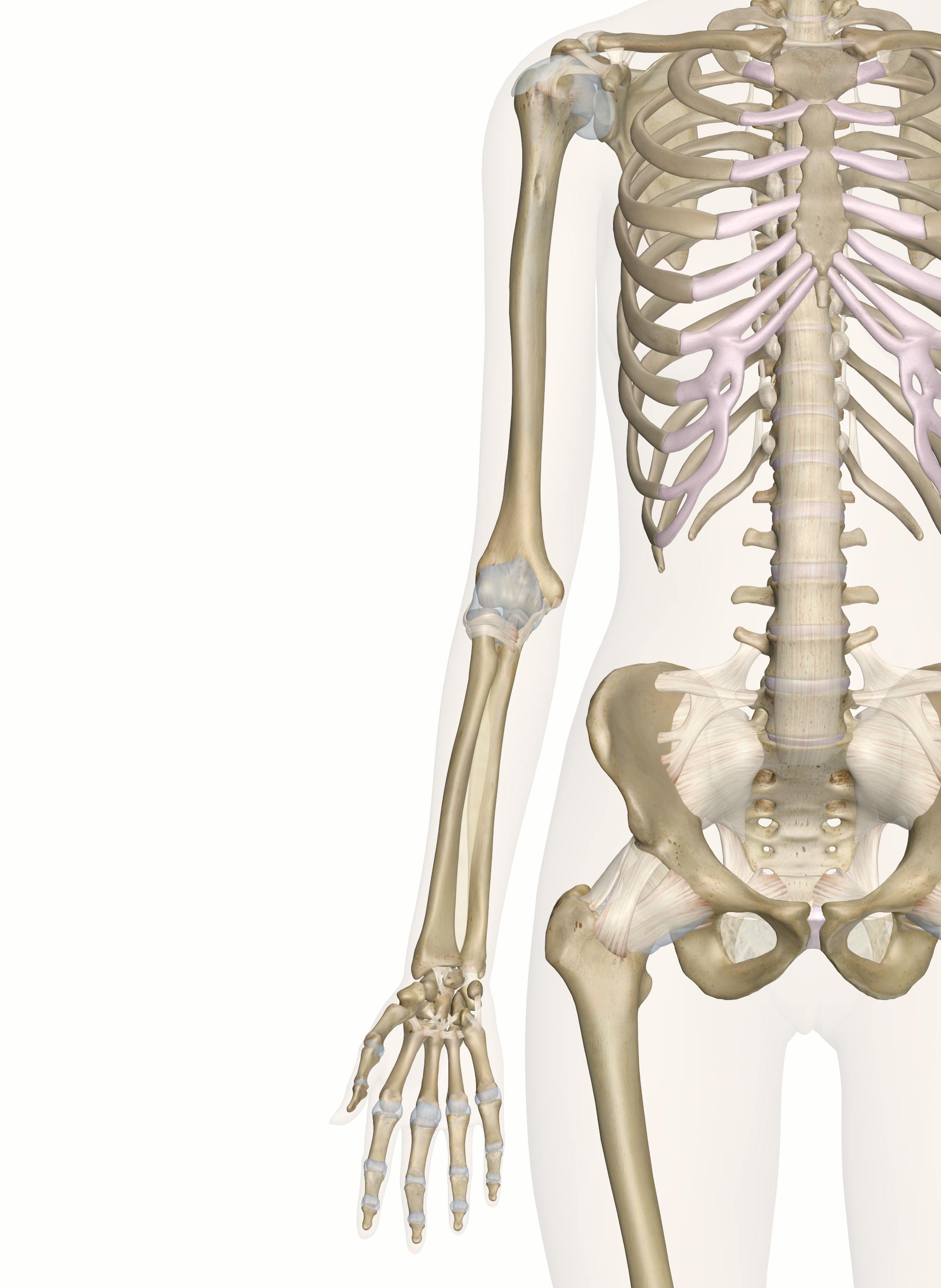
The last two pairs of ribs (11 and 12) are the smallest of all of the rib bones, and are called floating ribs. they get the name floating rib because they are connected to the spin at the back, but are not connected to anything at the front, thus appearing to float. Bone anatomy of head 12 photos of the bone anatomy of head bone anatomy of head, bone structure head office. The occiput (co), also known as the occipital bone, is a flat bone that forms the back of the head. Everything else that hangs from this, like the arms, legs, shoulders, and hips, is called the appendicular skeleton. The spine is composed of 33 bones called vertebrae, which stack together to form the spinal canal. This part of your anatomy is susceptible to injury, arthritis, herniated disks, pinched nerves and other problems. The normal curvature is 'outwards' (kyphosis). Browse 222 lower back skeleton stock photos and images available, or start a new search to explore more stock photos and images. The bones of the superior portion of the skull are known as the cranium and protect the brain from damage. Human left hand bone parts names There are seven cervical vertebrae, twelve thoracic vertebrae, and five lumbar vertebrae. Related posts of human back bone chart bone in arm pictures. Facet joints connect each vertebra, with fluid supporting.
Osteomyelitis (spinal bone infection) osteopenia (low bone mass density, not osteoporosis) osteoporosis topic center. Some individuals may also have additional (i.e., supernumerary) cervical ribs or lumbar vertebrae. The axial skeleton is made up of the skull, backbone, breastbone, and ribs. Sides of cranium, just above the ears. Vertebrae in these regions are essentially alike, with minor variation.
Anatomy Hand And Wrist Bid Needham from www.bidneedham.org The bones of the superior portion of the skull are known as the cranium and protect the brain from damage. At the sides of the cranium, higher than the temporal bones. Some individuals may also have additional (i.e., supernumerary) cervical ribs or lumbar vertebrae. Vertebrae, bones, joints, ligaments, muscles, muscular system, fascia, arteries, veins, nerves and various adjacent organs. The spine is composed of 33 bones called vertebrae, which stack together to form the spinal canal. For example, the sella turcica in the skull translates as turkish chair or turkish saddle. the image of a turkish chair—a backless chair with flaired, curling arms—is what this structure actually looks like. Back pain is one of the most common kinds of pain for adults, and muscle strains are the most common type. The hollow tube formed by the bony rings on the back of the spinal column surrounds the spinal cord.
A small bone in front of the temporal bone.
Back pain diagnosis often includes the name of the part of the spine from which your doctor believes the pain arises. Atlas (c1) the atlas is the first cervical vertebra and therefore abbreviated c1. The vertebral column of the lower back includes the five lumbar vertebrae, the sacrum, and the coccyx. The backbone is also called the vertebral column. Three curvatures of the vertebral column: On anatomical parts the user can choose to display the various structures in colored illustrations of the anatomy of the back and spine: It runs down the centre of the body. The spinal cord is like a long wire made up of millions of nerve fibers. There are 206 bones in the human body. Bone anatomy of head 12 photos of the bone anatomy of head bone anatomy of head, bone structure head office. The bones forming the dorsal part of the thoracic cage. Throughout the spine, intervertebral discs made of. Using this atlas of human anatomy of the spine and back.
Browse 222 lower back skeleton stock photos and images available, or start a new search to explore more stock photos and images. Bone in arm pictures 12 photos of the bone in arm pictures bone cancer arm pictures, pictures of bone cancer in arm, bone, bone cancer arm pictures, pictures of bone cancer in arm. These regions are called the cervical spine, thoracic spine, lumbar spine, sacrum, and coccyx. The axial skeleton is made up of the skull, backbone, breastbone, and ribs. An abnormal curve of the lumbar spine is lordosis, also called sway back.
Bones Of The Arm And Hand Interactive Anatomy Guide from innerbody.imgix.net An even smaller bone just to the sides of the eyes. Bone anatomy of head 12 photos of the bone anatomy of head bone anatomy of head, bone structure head office. These bones work together to provide flexibility to the trunk, support the muscles of the trunk, and protect the spinal cord and spinal nerves of the back. This part of your anatomy is susceptible to injury, arthritis, herniated disks, pinched nerves and other problems. The bones forming the dorsal part of the thoracic cage. Bone spurs that form on the facet joint can project into the tunnel, narrowing the hole and pinching the nerve. A small bone in front of the temporal bone. At the back of the cranium.
There are seven cervical vertebrae, twelve thoracic vertebrae, and five lumbar vertebrae.
The muscles, bones, ligaments, and tendons in the back can all be injured and cause back pain. The human vertebral column contains 33 vertebrae divided into seven cervical, 12 thoracic, five lumbar, five sacral and four coccygeal vertebrae, according to basic human anatomy. There are seven cervical vertebrae, twelve thoracic vertebrae, and five lumbar vertebrae. The most common variations include sutural (wormian) bones, which are located along the sutural lines on the back of the skull, and sesamoid bones which develop within some tendons, mainly in the hands and feet. Sides of cranium, just above the ears. The spinal cord is like a long wire made up of millions of nerve fibers. For example, the sella turcica in the skull translates as turkish chair or turkish saddle. the image of a turkish chair—a backless chair with flaired, curling arms—is what this structure actually looks like. Back pain diagnosis often includes the name of the part of the spine from which your doctor believes the pain arises. This vertebra supports the skull. The normal curvature is 'outwards' (kyphosis). This structure consists of bones called the vertebrae. Facet joints connect each vertebra, with fluid supporting. This section is made up of 12 vertebrae (abbr.
The bones of the superior portion of the skull are known as the cranium and protect the brain from damage back bones. This vertebra supports the skull.


0 Komentar