Bone Cross Section Microscope - Dinosaur bone cross section 972 x 1024 : ThingsCutInHalfPorn / New vs old newly designed microscope slide for cutting and viewing a quick cross section of textile fibers and small soft specimens of many types.
Bone Cross Section Microscope - Dinosaur bone cross section 972 x 1024 : ThingsCutInHalfPorn / New vs old newly designed microscope slide for cutting and viewing a quick cross section of textile fibers and small soft specimens of many types.. 1, cmp consists of both crystalline and glass phases fig. They build the entire picture, improve your understanding, consolidate the information and facilitate recall. Cross section performed on focused electon beam (fib) microscope at the university of kentucky's electron microscopy center. Monocots and dicots have distinct vascular bundles organization. Structural parts of a microscope and their functions.
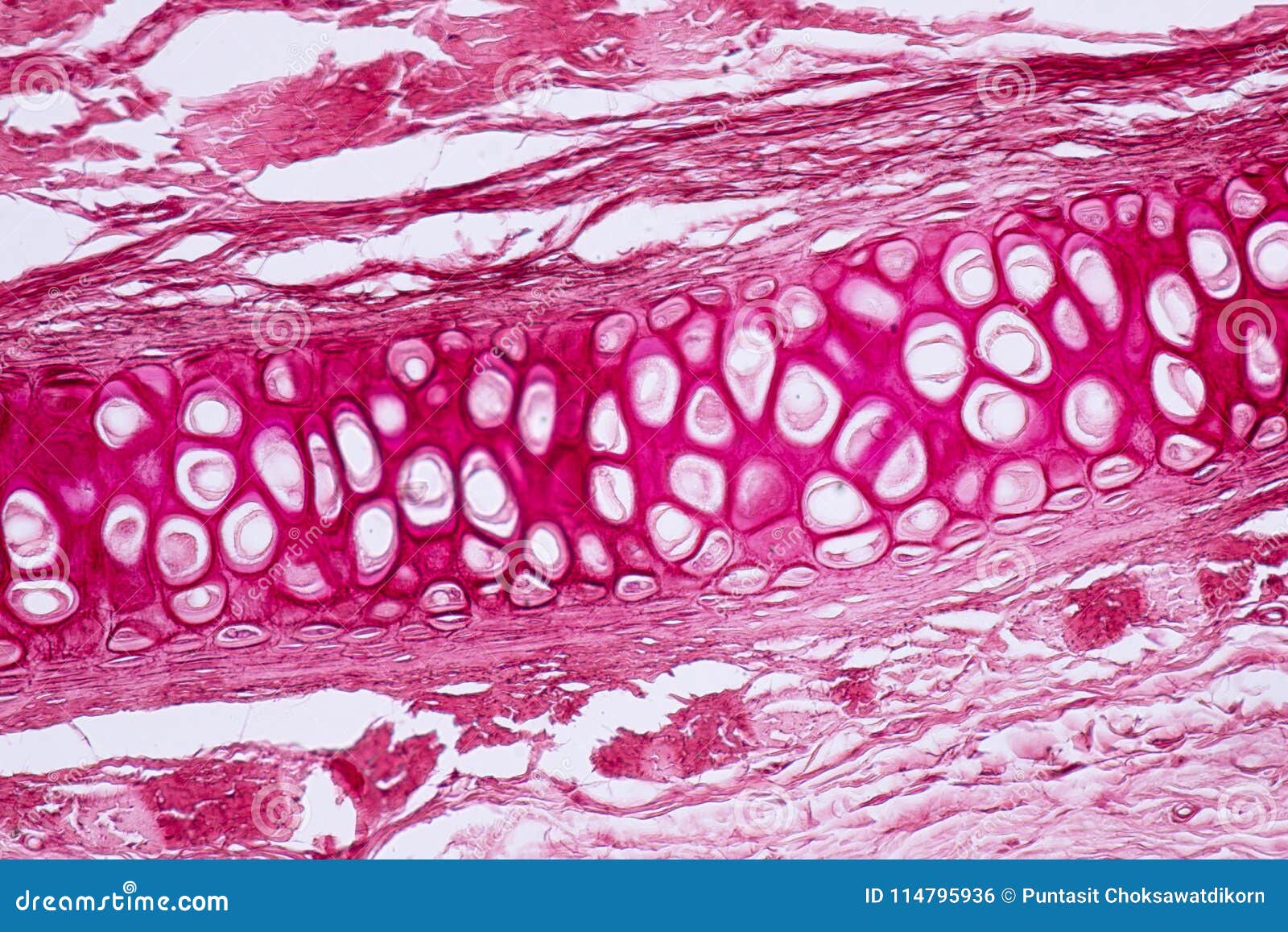
The microscopic cross section measures the probability of occurrence of a particular nuclear reaction. Jump to navigation jump to search. A cross section of a human long bone. Cross section bones medical mood google search health health care medicine med school. Use electromagnets to focus electrons resulting in significantly greater magnifications and resolutions.
Cross Section Human Cartilage Bone Under Microscope View ... from thumbs.dreamstime.com Structural parts of a microscope and their functions. This simply involves placing a section of the bone on the microscope stage and viewing. The microscopic cross section measures the probability of occurrence of a particular nuclear reaction. Cross section bones medical mood google search health health care medicine med school. Hi all, i have uploaded a new medical animation tutorial. They build the entire picture, improve your understanding, consolidate the information and facilitate recall. Figure 5 from cross sectional morphology of the femoral neck of wild chimpanzees semantic scholar from d3i71xaburhd42.cloudfront.net. File:earthworm crosssection stained microscope slide labeled.jpg.
Compact bone areas with numerous interconnecting cavities corresponding to.
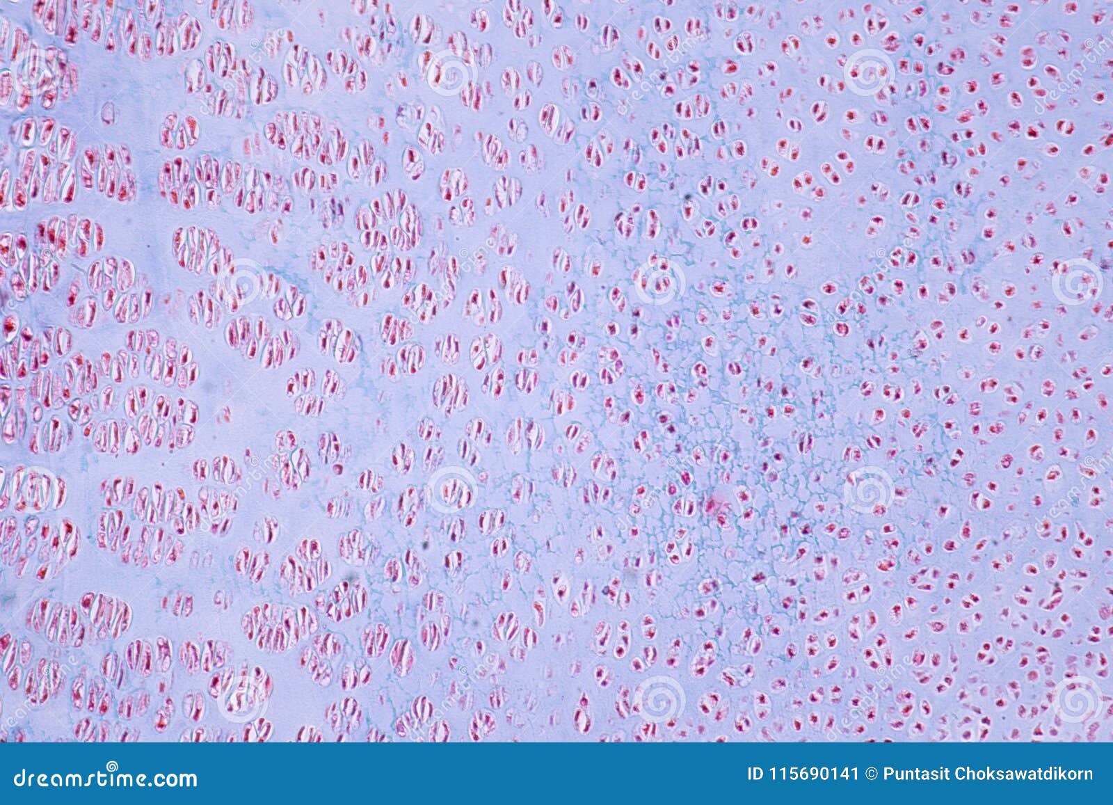
Bone marrow aspiration uses a hollow needle to remove a small sample (about 1 ml) of bone marrow for examination under a microscope. Compact bone cross section courtesy: A cross section of a compact bone shows concentric circles called lamellae. 1, cmp consists of both crystalline and glass phases fig. Important features in the bone cross section such as harvesian canals, osteons, osteon fragments, lamellar bone, bony trabeculae, myxoid matrix and artifact for. New vs old newly designed microscope slide for cutting and viewing a quick cross section of textile fibers and small soft specimens of many types. From wikimedia commons, the free media repository. Accuracy of the tested digitization method was expressed by. The microscopic bone cross section image acquired by using electronic microscope and is shown in fig.2. Thus as usual microscopic cross sections are experimentally measured. Both types of bone marrow are enriched with blood vessels and capillaries.2. See more ideas about microscopic, plant cell, microscopic. Monocots and dicots have distinct vascular bundles organization.
1, cmp consists of both crystalline and glass phases fig. These bone cells have long branching arms (d) which lets them communicate with. Cross section bones medical mood google search health health care medicine med school. New vs old newly designed microscope slide for cutting and viewing a quick cross section of textile fibers and small soft specimens of many types. Cross section of ground compact bone.
Dinosaur Bone Cross-Section Under the Microscope ... from i.pinimg.com This is a short tutorial using blender 2.8 that shows how to create a bone cross section and using images to create the textures. The large dark spots are passages for blood vessels and nerves. Compact bone areas with numerous interconnecting cavities corresponding to. From wikimedia commons, the free media repository. Accuracy of the tested digitization method was expressed by. Sometimes referred to as 'spongy bone' or 'trabecular bone', cancellous bone is found within the middle of large bones. The microscopic bone cross section image acquired by using electronic microscope and is shown in fig.2. In this short video i use blender 2.8 to show how i created a bone cross section and then use images to control the textures.
Bone marrow aspiration uses a hollow needle to remove a small sample (about 1 ml) of bone marrow for examination under a microscope.
Accuracy of the tested digitization method was expressed by. 1, cmp consists of both crystalline and glass phases fig. Thin section of dinosaur bone. A cross section of a human long bone. Use electromagnets to focus electrons resulting in significantly greater magnifications and resolutions. This is a short tutorial using blender 2.8 that shows how to create a bone cross section and using images to create the textures. Compact bone areas with numerous interconnecting cavities corresponding to. Hope you enjoy and please. The microscopic bone cross section image acquired by using electronic microscope and is shown in fig.2. When the light that enters the condenser is polarized by placing a polarizer in the filter holder and a second, crossed polarizer at the image plane. See more ideas about microscopic, plant cell, microscopic. The concept of a nuclear cross section can be quantified physically in terms of characteristic area where a larger area means a larger probability of interaction. Cross section performed on focused electon beam (fib) microscope at the university of kentucky's electron microscopy center.
See more ideas about microscopic, plant cell, microscopic. The finished bone section will be bonded to a microscope slide and so the first step is to grind flat and polish the part of the bone that will be glued to the slide. Monocots and dicots have distinct vascular bundles organization. Thin section of dinosaur bone. Microscope cross section (page 1).
Cross Section Human Cartilage Bone Under Microscope View ... from thumbs.dreamstime.com This is a short tutorial using blender 2.8 that shows how to create a bone cross section and using images to create the textures. In this short video i use blender 2.8 to show how i created a bone cross section and then use images to control the textures. From wikimedia commons, the free media repository. The finished bone section will be bonded to a microscope slide and so the first step is to grind flat and polish the part of the bone that will be glued to the slide. Hope you enjoy and please. Cross section bones medical mood google search health health care medicine med school. See more ideas about microscopic, plant cell, microscopic. This simply involves placing a section of the bone on the microscope stage and viewing.
This is a short tutorial using blender 2.8 that shows how to create a bone cross section and using images to create the textures.
A cross section of a compact bone shows concentric circles called lamellae. Scanning electron micrograph scanning electron microscope images microscope pictures microscopic photography microscopic images cross section macro and micro human anatomy. Cross section performed on focused electon beam (fib) microscope at the university of kentucky's electron microscopy center. This simply involves placing a section of the bone on the microscope stage and viewing. When the light that enters the condenser is polarized by placing a polarizer in the filter holder and a second, crossed polarizer at the image plane. Both types of bone marrow are enriched with blood vessels and capillaries.2. New vs old newly designed microscope slide for cutting and viewing a quick cross section of textile fibers and small soft specimens of many types. Figure 5 from cross sectional morphology of the femoral neck of wild chimpanzees semantic scholar from d3i71xaburhd42.cloudfront.net. 1, cmp consists of both crystalline and glass phases fig. Thus as usual microscopic cross sections are experimentally measured. Structural parts of a microscope and their functions. Sometimes referred to as 'spongy bone' or 'trabecular bone', cancellous bone is found within the middle of large bones. These bone cells have long branching arms (d) which lets them communicate with.
Important features in the bone cross section such as harvesian canals, osteons, osteon fragments, lamellar bone, bony trabeculae, myxoid matrix and artifact for bone cross section. In this short video i use blender 2.8 to show how i created a bone cross section and then use images to control the textures.


0 Komentar